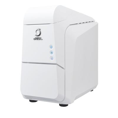
JEOL NeoScope JCM-7000 Benchtop SEM


Benchtop scanning electron microscopes are used in a wide range of fields, such as electrical, electronics, automobiles, machinery, chemical, and pharmaceutical industries. In addition, SEM applications are expanding to not only cover research and development, but also address quality control and product inspection at manufacturing sites. With this, demands for further improved work efficiency, much faster and easier operation, and a higher degree of analytical and measurement capabilities, are increasing.
The JCM-7000 Benchtop Scanning Electron Microscope is designed based on a key concept of "Easy-to-use SEM with seamless navigation and live analysis". The JCM-7000 incorporates three innovative functions; "Zeromag" for smooth transition from optical to SEM imaging, "Live Analysis" for finding constituent elements for an image observation area, and "Live 3D" for displaying a reconstructed live 3D image during SEM observation.
When you place the JCM-7000 next to an optical microscope, further-faster and more-detailed foreign material analysis and quality control can be made.
Features:
- Zeromag - simplifies navigation and enhances throughput. Provides a seamless transition from an optical (or holder graphic) to SEM image
- Highly-advanced Auto functions for automatic condition setting and image formation in minutes
- High resolution (100,000X) and large depth of field
- High and low vacuum modes for managing a wide variety of samples
- Large chamber : maximum sample size 80mm (D) x 50mm (H)
- Advanced functions built-in such as : Automated montage and Live 3D imaging
- Option: Fully embedded EDS with Live (real-time) analysis
- Smile ViewTM Lab for integrated management of image and analysis data
ZEROMAG
- Zeromag simplifies navigation and enhances throughput
- With Zeromag, you can navigate from a colour optical image - as you increase the magnification, you transition from the optical to live SEM image automatically
- Set up large area automated image monatge and stiching; EDS option includes automated montage X-ray map
- Automatically link SEM image, position, optical image, and EDS data (with EDS option)
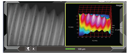
LIVE 3D IMAGING
 With the EDS option, the 6-channel, high sensitivity, solid state backscatter electron detector acquires composition, topographic and shadow (combination of composition and topography) images, and supports live 3D imaging.
Combine the live 3D image with software to create a 3D model and calculate surface texture data including cross-sectional profile, height and surface roughnesss
LIVE ELEMENTAL ANALYSIS
Utilising the optional EDS, with live elemental analysis, you can:
- View EDS- spectra in real time as you search for an area of interest
- Set analysis points, area, map positions and line scans
- View major elements detected, and automatically display on live EDS window
For further information please contact us or download the datasheet.
| Mode | High-vacuum mode: Secondary electron image Backscattered electron image (Composition, topographic and shadow. 3D images) Low-vacuum mode: Backscattered electron image (Composition, topographic and shadow, 3D images) |
| Electron gun | Tungsten filament/Wehnelt integrated grid |
| Accelarating voltage | 3 stages 15kV/10kV/5kV |
| Specimen stage | X-Y motor drive stage X: 40mm; Y: 40mm |
| Maximum specimen size | Diameter 80mm, height 50mm |
| Specimen exchange | Draw-out mechanism |
| Pixels for image acquisition | 640 x 480 1280 x 960 2,560 x 1,920 5,120 x 3,840 |
| Automated functions | Alignment, focus, stigmator, brightness/contrast |
| Measurement functions | Distance between 2 points, angles, line wdith |
| File format | BMP, TIFF, JPEG, PNG |
| Computer | Desktop PC Windows® 10 |
| Monitor | 24 inch |
| Vacuum system | Full-automatic TMP:1, RP:1 |

