News
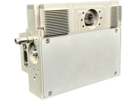
New Laser Driven Light Source
Energetiq have built upon the success of previous models to develop their next-generation light source, the EQX-850 LDLS. Its innovative design offers enhanced performance, user experience, and value to meet the tough demands of the leading-edge semiconductor processes.
Features and benefits:
- Ensures uniform results across all tools and processes.
- Delivers unrivaled short-term stability and higher spectral radiance.
- Features a modular design for easy maintenance and eliminates the need for water cooling.
- Includes event logging, remote software interface controls, and performance monitoring.
For further information please contact us or read more.
Read MoreEDAX Octane Elite Ultra EDS System
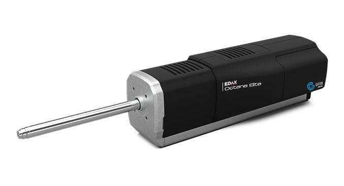
Gatan, Inc., a business of AMETEK Inc and a global leader focused on enhancing and extending the operation and productivity of electron microscopes, has launched the EDAX Octane Elite Ultra energy dispersive x-ray spectroscopy (EDS) system for scanning electron microscopes.
The Octane Elite Ultra EDS establishes a new benchmark for EDS systems by introducing a proprietary windowless 160 mm2 detector. This detector delivers superior light and heavy elements sensitivity while providing accurate analytical results at all accelerating voltages.
"For many years, researchers needing extreme sensitivity in their elemental analysis have been forced to forego accurate analytical results or invest in additional detectors. Due to interference from other signals in the microscope, EDS detectors optimized for sensitivity permitted qualitative investigations only," said Dr. David Stowe, Senior Product Manager, Gatan. "Combining a large active area and harnessing the power of an electron trap to prevent pollution of the signal, the Octane Elite Ultra makes compromise a thing of the past."
"With an active area >80% larger than other detectors and an ability to provide precise quantitative results, the Octane Elite Ultra is the first EDS detector that provides the cutting edge of research while also permitting routine analytical measurements," commented Narayan Vishwanathan, Vice President and Business Unit Manager of AMETEK Electron Microscopy Technologies. "It eliminates the need to switch between detectors for different measurements, allowing users to focus on the most important thing: their research."
For further information please contact us or read more.
Read MoreNew Microscopes, Enhanced Optical Performance
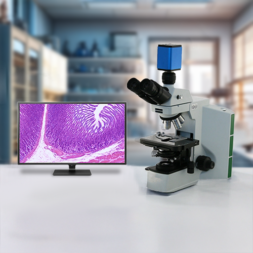
WPI is excited to announce the launch of a new line of microscopes featuring cutting-edge optics and advanced light sources designed for enhanced visibility and precision. These innovative instruments utilise high-resolution lenses and state-of-the-art illumination technologies, ensuring that users can capture even the finest details in their specimens. Whether you're in a research lab, educational setting, or industrial environment, these microscopes are tailored to meet diverse needs.
LCD Laboratory Microscopes
Inverted Teaching Microscopes
For further information please contact Tim Watts.
Read MoreNew Optical Filters with Improved Edge Steepness
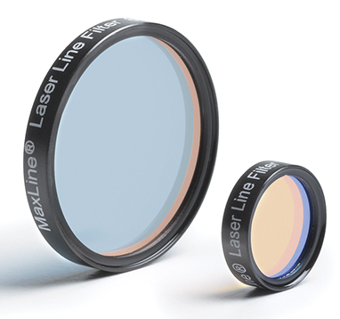
The new MaxLine E-Grade optical filters from Semrock offer improved and superior edge steepness for improve blocking of the laser line, and reduced Rayleigh-scattering.
In combination with a RazorEdge filter, these make the ideal pair for Raman Spectroscopy. The MaxLine filter spectrally "cleans up" the excitation laser light before it reaches the sample under test, allowing only the desired laser line to reach the sample. The RazorEdge filter then removes the laser line from the light scattered by the sample, while efficiently transmitting desired light at wavelengths very close to the laser line.
The initial release of the E-Grade MaxLine laser line filters will include version for 488nm, 532nm, 633nm and 785nm.
For further information please contact Julia King or read more.
Read MoreNew Performance Enhanced Marana BI sCMOS Camera
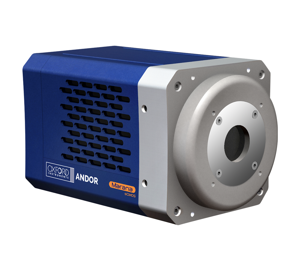
Oxford Instruments Andor, a world leader in scientific imaging solutions, has today announced the launch of a performance-enhanced back-illuminated sCMOS camera, further strengthening its broad portfolio of cameras for Physical Sciences and Astronomy.
The performance of the Marana 4.2B-6 back-illuminated 4.2 Megapixel sCMOS model has been significantly enhanced to widen its application appeal within Physical Sciences and Astronomy. A new Low Noise Mode reduces the read noise to 1.0e-. When combined with market-leading -45°C vacuum cooling and 95% QE, this pushes the limits of detection further, even under the most challenging, light starved imaging applications, enabling tracking of smaller Space Debris or NEOs, shorter exposures, lower illumination powers to protect photosensitive samples or the detection of trace concentrations of species.
A new High-Speed mode has been implemented to meet the needs of fast imaging applications such as trapped ion/atom quantum computing, solar astronomy, fast spectroscopy or hyperspectral imaging. By combining this mode with a 2-lane CoaXPress connection, 135 fps of sustained and stable high-speed operation is now possible.
Furthermore, a new Long Exposure Mode has been implemented which markedly enhances the exposure flexibility of Marana 4.2B-6. Amplifier glow has been a problem that has plagued most sCMOS sensors on the market. This new mode goes a long way to suppressing the effect of amplifier glow under longer exposure conditions. This is particularly relevant for fields such as astronomy and low light luminescence detection.
A ‘Global Clear’ mode has now been implemented for the Rolling Shutter sensor type. This mode purges charge from all rows of the sensor simultaneously at the exposure start. It can be used alongside a pulsed/triggerable light source, such as LED or Laser, to simulate a Global Shutter mechanism, useful for achieving tight synchronisation with other equipment and minimising exposure 'dead times'.
For further information please contact Michael Buckett, or read more.
Read More