News
Webinar : Compact and Upgradeable Benchtop Fluorescence Microscope
Date : Wednesday 27 November 2024
Time : 5am NZDT, 3am AEDT, Midnight AWST
Speaker : Dr Geraint Wilde
If you are not a night owl, please register and a recording will be emailed to you
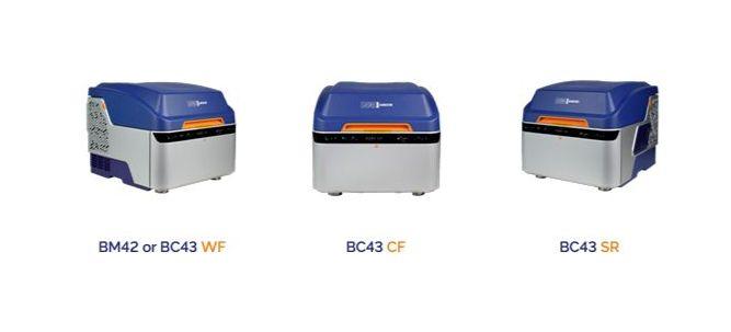
Andor is excited to announce the expansion of our Benchtop Microscope range, adding application and budget-based flexibility to the original BC43 Benchtop Confocal. Their Benchtop Microscopes now start with motorised multi-dimensional widefield fluorescence imaging and extend into super-resolution range.
They will introduce the new products and the different imaging modalities of widefield, confocal and super-resolution fluorescence microscopy. The biological models and typical experiments those modalities can be optimally used with will be discussed.
Being a modular design, they will cover how our latest products can be upgraded in the field over time as your research needs change.
Learning Objectives
- Learn about the new extended BC43 product range
- Gain Insight into different imaging modes (widefield, confocal and super-resolution) and the sample types they are applicable to
- Understanding the achievable multiple scales of imaging
Read More
Webinar : Performing Accurate Force Measurements on Biological and Soft Matter
 Date: Wednesday 23 October
Time: 2am AEDT, 4am NZDT, 11pm AWST

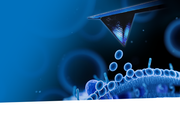
Join us and special guest speaker Prof. Andreas Janshoff from Georg-August University Göttingen, Germany, for this webinar on performing accurate force measurements on biological and soft matter using atomic force microscopy (AFM).
Forces play a crucial role in biological mechanisms, such as cellular response, molecular interactions, and protein binding, and are essential for deriving the nanomechanical properties of a sample. AFM has emerged as a key platform for the precise measurement of interaction forces on the nanometer scale. AFM force spectroscopy is used to quantify forces and determine nanomechanical properties such as Young's modulus, cell adhesion, and viscoelastic properties, information that is invaluable for studying interaction-based and disease-related biomechanical changes.
Prof. Janshoff will outline his work using AFM to investigate the biomechanical properties of cell membranes, and, in particular, the viscoelastic behavior of the cell cortex.
Webinar Highlights:
- Key insights and proven strategies for achieving reliable results
- Common pitfalls and practical tips for ensuring reproducible measurements
- Q&A session with the team
New Microscopes, Enhanced Optical Performance
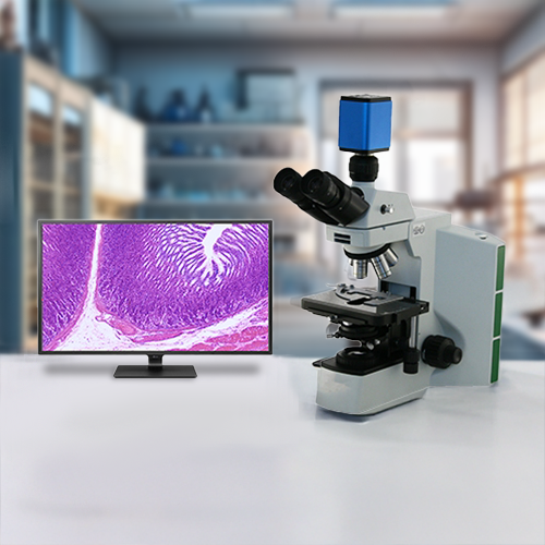
WPI is excited to announce the launch of a new line of microscopes featuring cutting-edge optics and advanced light sources designed for enhanced visibility and precision. These innovative instruments utilise high-resolution lenses and state-of-the-art illumination technologies, ensuring that users can capture even the finest details in their specimens. Whether you're in a research lab, educational setting, or industrial environment, these microscopes are tailored to meet diverse needs.
LCD Laboratory Microscopes
Inverted Teaching Microscopes
For further information please contact Tim Watts.
Read MoreCompact and Upgradeable Benchtop Fluorescence Microscope
Date: Wednesday 19 June 2024
Time: 3am NZST, 1am AEST, 11am AWST
If you are not a night owl register for the webinar and you will receive a copy after the event :)
With the latest expansion of our Benchtop Microscope range, adding application and budget-based flexibility to the original BC43 Benchtop Confocal. We can now offer more choice and scales of imaging for Neuroscience studies. Our Benchtop Microscopes now start with motorised multi-dimensional widefield fluorescence imaging and extend into super-resolution range.
We will introduce our new products and the different imaging modalities of widefield, confocal and super-resolution fluorescence microscopy. The typical biological models and experiments those modalities can be optimally used with will be discussed.
Learning Objectives
- Learn about the new extended BC43 product range
- Insight into different imaging modes (widefield, confocal and super-resolution) and how they can be applied to typical neuroscience research models and experiments.
- Understanding the achievable multiple scales of imaging.
Geraint Wilde attained a Ph.D. in Neuroscience in 1997 from the University of Southampton, UK, and then continued in the field with a postdoctoral position at the University of Warwick, UK. Having developed an interest in microscopy during his Ph.D. and post doc, he moved to the University of Liverpool, UK, to work in the laboratory of Michael White, focusing on intracellular signalling and gene expression through live-cell imaging. Geraint then left academia to pursue a commercial career in microscopy, starting as a Sales Application Specialist for general life science microscopy where he was exposed to an even broader range of microscope technology and most importantly applications. He joined Andor Technology in 2009, where he is now Hardware Business Manager for Microscopy and Life Science Cameras and the Product Manager for the Andor Benchtop Microscopes.
Read MoreExpanding the Dragonfly Series
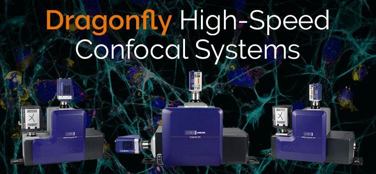
Oxford Instruments Andor, a world leader in scientific imaging solutions, today announces the launch of Dragonfly 400 and new VLE (Versatile Laser Engine), further expanding the award-winning Dragonfly confocal microscopy portfolio.
The Dragonfly 200 series offers speed, sensitivity and excellent confocal imaging, making it an ideal system for a wide range of applications, including live cell imaging, development biology, neurobiology, and cancer research. The Dragonfly 200 delivers high-performance spinning disk confocal imaging of all (thick, thin, fixed and live) biological samples. High-resolution image stacks are ready for analysis in mere seconds.
The upgraded model adds new capabilities including the new VLE (Versatile laser engine), which allows seven laser lines in a single chassis and up to 10 lines in dual chassis mode, giving researchers the capability to choose more fluorophores for their experiments as well as increasing labelling options on a single experiment. Furthermore, the Dragonfly series is built with modularity in mind, allowing Dragonfly 200 systems to be upgraded to the new Dragonfly 400 model in the field.
The new Dragonfly 400 series builds on the key features of the Dragonfly 200 by adding the established HLE (High Power Laser engine) and the 3D-Super Resolution Module. With the Dragonfly 400 series and the high-power laser engine (HLE), the system becomes ideal for an even wider set of research applications. The HLE delivers enough power for super-resolution applications (SMLM) with DNA-PAINT. A single plane image through the 3D super-resolution module delivers axial information over an ~1 μm range with a corresponding axial resolution down to 30 nm. The high-power lasers of the HLE deliver an increase in productivity over the Dragonfly 200 series, allowing even higher productivity in time-consuming applications, such as spatial omics applications and large sample imaging.
For the ultimate range of imaging applications, users have the option of choosing the Dragonfly 600 series with Borealis-TIRF and Zoom Illumination optics imaging functionality to make a complete multi-modal system for widefield, Confocal, TIRF, and SMLM (DNA-PAINT & dSTORM) applications. Dragonfly 600 has achieved impressive resolutions down to 6 nm (DNA-PAINT) and allows SMLM in any imaging modality (confocal, widefield, or B-TIRF).
For further information please contact Mark Richardson or read more.
Read More


