Bruker CellHesion 300 AFM
Bruker CellHesion 300 AFM
Automated, quantitative single-cell and tissue mechanics
![]()
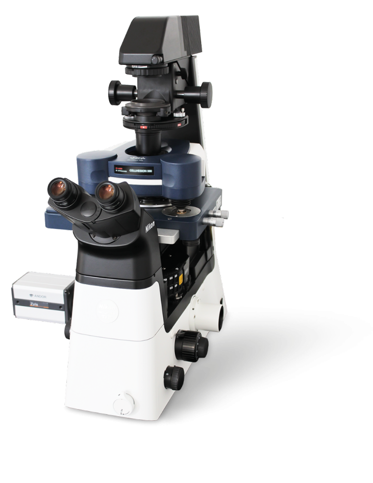
The automated CellHesion® 300 is the ideal tool for measuring cell-cell, cell-tissue, and cell-substrate interactions with single-molecule sensitivity. It enables fast and easy measurement of the structure, morphology, and nanomechanical properties of living biological systems, delivering crucial insights into the role they play in various pathological disorders. This innovative system creates novel possibilities for applications in biophysics, biochemistry, implant research, wound healing, developmental biology, stem cell research, infection biology and immune response studies.
CellHesion 300 provides reproducible, quantitative data of unprecedented quality. Its high degree of automation increases throughput and delivers the productivity, performance, and statistical significance required for biomedical and clinical research environments. Important parameters, such as maximum adhesion force, individual unbinding events, and tether characteristics are automatically determined from each dataset by innovative software solutions.
Only CellHesion 300 delivers:
- Maximised throughput for higher productivity by automating measurements
- Correlative and label-free multiparametric, nanomechanical measurements of living samples in near-physiological conditions
- Fast and simple selection of regions of interest over large sample areas, ideal for tissue biopsies
- Systematic workflow with visual support
For further information please contact us or download the datasheet.
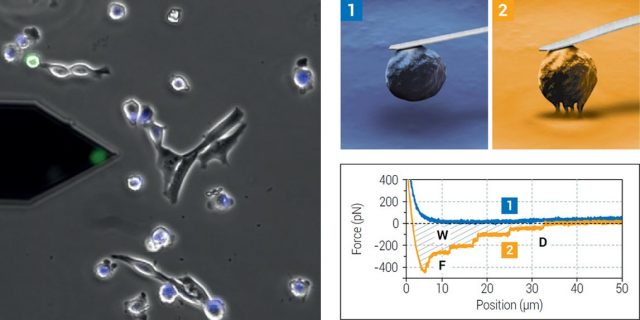
Left: Probe with a single 3T3 fibroblast (green, FDA staining) attached for cell adhesion measurements on a substrate or target cells. Right: In a Single Cell Force Spectroscopy (SCFS) experiment, a single living cell is biochemically bound to a probe (e.g., via functionalisation). The cell is brought into contact with the binding target and a defined force applied to the cell (1). After a user-defined binding period, the cell is separated from target by retracting the probe (2). The resistance to separation is measured by quantification of the probe deflection.
Cellesion 300 brochure Read more on Bruker's website