Bruker NanoWizard PURE BioAFM

Bruker NanoWizard PURE BioAFM
Streamlined design with best-in-class performance and flexibility

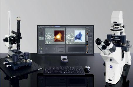
The NanoWizard PURE atomic force microscope combines functionality and performance in a high-value instrument. Based on the renowned NanoWizard technology platform, it delivers high-resolution imaging and nanomechanical analysis capabilities that can be seamlessly combined with advanced optical microscopy techniques.
Renowned quality
NanoWizard PURE delivers state-of-the-art functionality:
- Unique 3D tip-scanning with capacitive sensor technology
- PeakForce Tapping and QI modes for high-resolution imaging and advanced force control on delicate samples
- Intuitive operation and automated routines, analysis, and data processing capabilities provided by latest V8 software environment
Unrivalled excellence in its class
Equipped with innovative hardware and software features, the NanoWizard PURE is ideal for laboratories wishing to expand their research capabilities with cutting-edge correlated microscopy techniques, whether for experienced users or those new to AFM.
The unparalleled modularity of the NanoWizard platform provides outstanding versatility. Its easy handling and user-friendliness make it deal for multi-user imaging facilities.
- Discover attractive default configurations for biological and standard applications that offer best value solutions and capabilities
- Keep apace of technological advances with new modules and features that match your research requirements
- Upgrade paths to Bruker BioAFM premium NanoWizard product lines
Superior flexibility
NanoWizard PURE provides innovative research capabilities across a range of scientific fields, from the investigation of highly delicate biological samples, living cells, single molecules, and tissues, to the quantification of nanomechanical properties and study of polymers, soft matter, and advanced materials.
For further information please contact us or download the brochure.
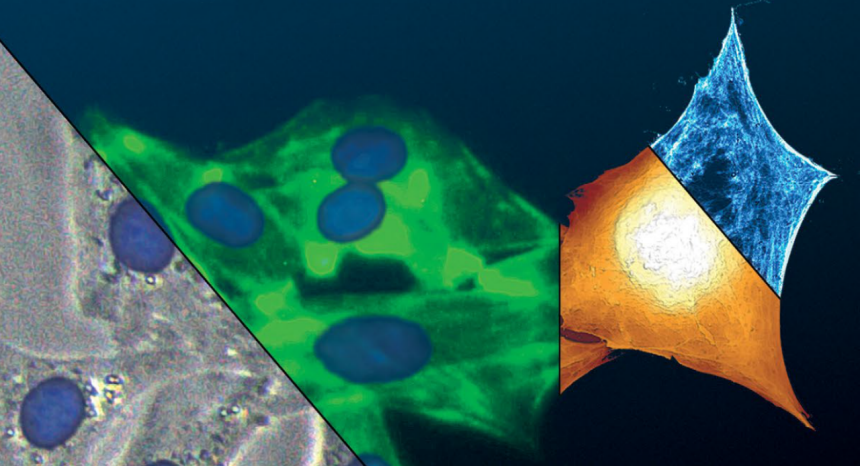
Correlated QI and optical images of live 3T3 fibroblast cells in cell culture medium at 37°C. Cell nuclei and actin fluorescently labelled with Hoechst 33342 and CellMask™ Green Actin Tracking, respectively. DirectOverlay semi-transparent superimposition of fluorescence images with optical phase contrast and QI scan. Scan size: 50μm × 70μm. Height range: 4μm (brown); Young’s modulus range: 20kPa (blue). Sample courtesy of Dr. Wedepohl, Freie Universität Berlin, Germany.
